
Seizures, Abdominal Pain and PRES Could Indicate AIP, Reports Suggest


Patients with abdominal pain, seizures and brain imaging indication of posterior reversible encephalopathy syndrome (PRES) could have acute intermittent porphyria (AIP), according to two new case reports and a literature review.
The study, “Acute intermittent porphyria presenting with seizures and posterior reversible encephalopathy syndrome: Two case reports and a literature review,” appeared in the journal Medicine.
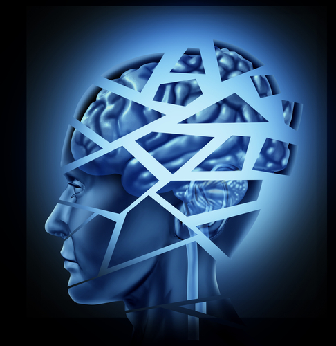
PRES patients experience headaches, seizures, visual disturbances, and/or mental dysfunction. AIP, caused by deficient biosynthesis of heme — a component of several iron-containing proteins such as hemoglobin, the protein that carries oxygen in the blood — presents in various ways, the most common being an acute attack with severe abdominal pain and nervous system disturbances.
Seizures and hallucinations were first reported in 1991 in an AIP patient who also showed reversible lesions on a brain magnetic resonance imaging (MRI) scan. Since then, a growing number of suggestive PRES cases in patients with AIP have been reported. However, the link between PRES and AIP still needs further study.
A team from China presented two AIP cases with seizures and PRES, and discussed 20 published cases (20 women, average age at AIP onset 23) to raise awareness of the importance of early and accurate diagnosis.
Case one refers to a 27-year-old man hospitalized for abdominal and lumbar pain lasting 12 days after heavy food and alcohol intake. He had been diagnosed with acute pancreatitis with mild elevated urine amylase, an enzyme made by the pancreas that helps break down starch. His blood pressure then increased, followed six days later by a generalized tonic-clonic seizure, which involves the entire brain, and partial seizures in the left limb.
The patient had an unremarkable clinical examination, except for decreased tendon reflex — nerve reflexes that determine muscle contraction upon tapping — in the four limbs. He also exhibited tachycardia, or faster heartbeat.
Biochemical evaluation showed elevated levels of creatine kinase — which may indicate heart attack, skeletal muscle injury, or heavy drinking — and aspartate transaminase — a marker of liver damage — while serum sodium was reduced (hyponatremia).
His brain MRI scans revealed multifocal lesions. Further analysis indicated vasogenic edema, which refers to a breakdown of the blood-brain barrier. His recurrent abdominal pain without clear cause was suspect of AIP. The diagnosis of AIP was made after three positive urine Watson-Schwartz tests, in which his urine turned dark and red upon exposure to light.
The patient responded well and recovered gradually with glucose infusion and a high-carbohydrate diet. Treatment with hematin (a heme derivative) was not available in the hospital. He was later discharged with no seizures or abdominal pain, remaining asymptomatic afterwards.
Case two refers to an 18-year-old girl hospitalized with generalized tonic-clonic seizures. She recently had been diagnosed with ileus (lack of intestinal movement) as the result of abdominal pain and constipation. An abdominal X-ray, computerized tomography (CT) scan, and ultrasound were normal.
The patient’s MRI scan indicated PRES, but the diagnosis was unclear. Because of seizures, she was transferred to the emergency department. She was then given anticonvulsants including benzodiazepines and oxcarbazepine. The clinicians suspected AIP based on recurrent abdominal pain and seizures. She had no family history of similar symptoms.
Her blood pressure and heart rate were high. She experienced a hallucination on the night of admission. She had marked hyponatremia — possibly involved in the occurrence of seizures — low serum potassium, and high aspartate transaminase, alanine transaminase and total bilirubin, also indicators of liver damage.
Six days later, a brain MRI revealed partial lesion resolution. AIP diagnosis was confirmed by three Watson-Schwartz tests. She was treated with a high-carbohydrate diet and intravenous dextrose, identical to glucose. Upon improvement, she was discharged with no seizures and less abdominal pain.
An MRI two weeks later confirmed the disappearance of brain lesions. One month later, she had recovered and gone back to work. No acute attacks subsequently occurred.
As for the literature review, the investigators found that most AIP attacks occurred in a patient’s 20s or 30s and the mean age at PRES presentation was 44 years. However, both in the two patients they described and the cases they reviewed, the scientists observed earlier onset when AIP and PRES occurred together.
Seizures during acute AIP attacks were reported in all but one reviewed cases. “Taken together, seizures are likely the most prominent dysfunction of nervous system during acute attack of AIP patients with PRES,” the team wrote.
Unlike headache, constipation, visual impairment, neuropsychiatric symptoms — including hallucination, drowsiness and confusion — and, particularly, abdominal pain were common in AIP patients with PRES. Hypertension, tachycardia and hyponatremia were also frequent. Most reviewed studies indicated vasogenic edema as a possible mechanism.
“AIP should always be suspected in the setting of MRI presentation of PRES in patients with unexplained abdominal pain and seizures,” the investigators said.